
Despite their extensive fossil record, the skeletal structure of early jawed vertebrates remains poorly documented. This project will investigate the mechanics of bone remodelling and growth in the head and trunk dermal skeleton of some of the earliest osteichthyans. These data will be placed in a phylogenetic context by comparison with ancestral states in extinct groups of jawed vertebrates ('placoderms').
Backgroundand context
An external skeleton of skin 'teeth' and bony plates is a major evolutionary adaptation of vertebrates that primitively appeared in fossil groups of heavily 'armoured' jawless fish. The competence to extensively resorb and modify dermal bone, however, originated in primitive jawed vertebrates ('placoderms') and is also a feature of modern osteichthyans. How skeletal plasticity was maintained in these early lineages compared to model vertebrate species remains an open question.
During this study you will sample 'placoderm' and osteichthyan species from the middle Palaeozoic (425-420 million years ago) for evidence of developmental changes to the structure of the dermal skeleton. On this basis, you will interpret growth pattens in the dermal skeleton of these taxa and explore the influence of developmental characters on the phylogenetic tree of early vertebrates.
Objectives and goals
This internship project has three main aims:
- Record the structural and tissue composition differences between the dermal skeleton of 'placoderms' and early osteichthyans.
- Identify the factors responsible for observed changes in bone structure (e.g. mechanical stress, tissue renewal or tissue growth)
- Produce a phylogenetic hypothesis for the evolution of dermal bone in early jawed vertebrates using the new developmental data.
Materialsand methods
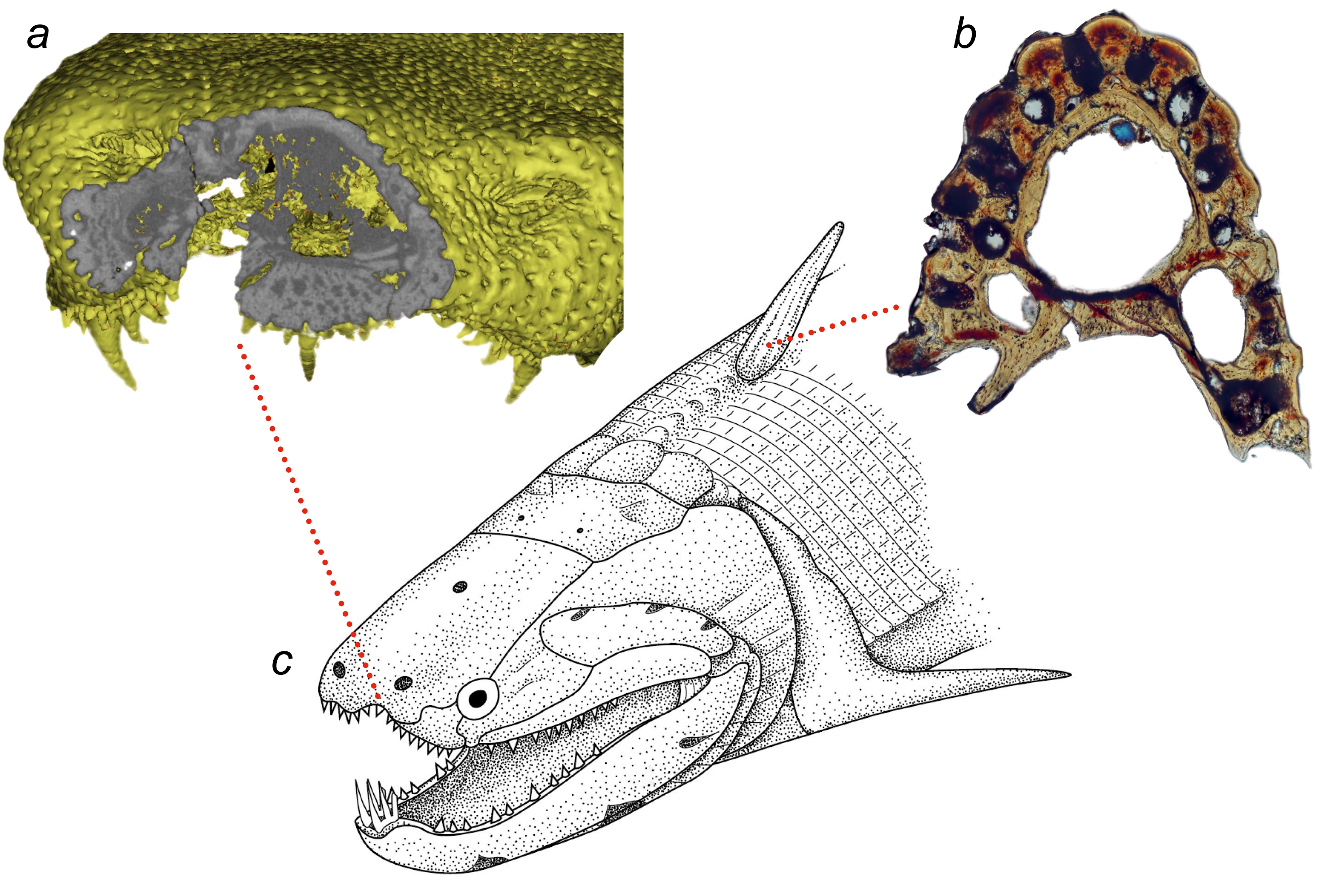
During the project you will examine fossil material from south China representing dermal scales and fin spines, as well as head and shoulder girdle plates, from the upper Silurian 'placoderms' Entelognathus and Qilinyu and the Lower Devonian osteichthyans Psarolepis and Youngolepis. The specimens will be analysed with high resolution micro-CT tomography and Nomarski polarised light microscopy.
Studentrequirements
Under supervision you will collect and analyse tomography and light microscopy image data from fossils.
Basic knowledge of vertebrate anatomy and skeletal tissues as well as some experience in 3D rendering and/or digital arts are desirable for the project.